
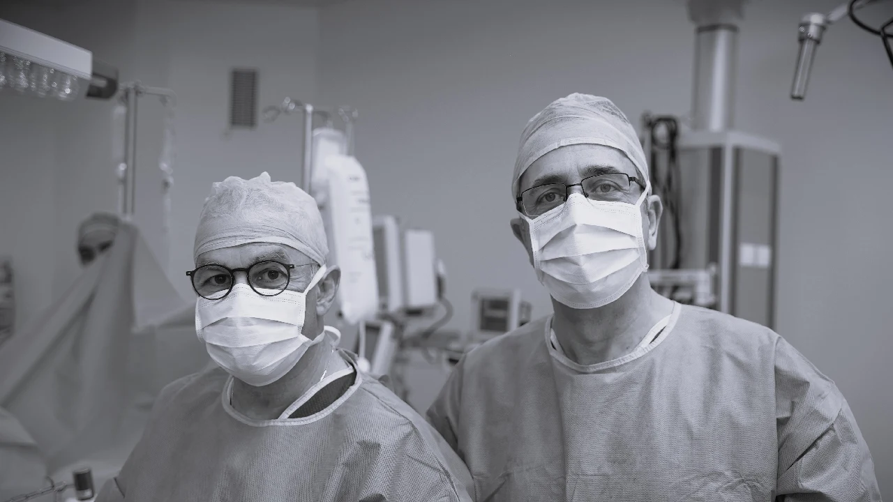
Abdominal sarcomas: why they are difficult to operate on
Abdominal sarcomas are difficult to operate on. They are usually large when diagnosed and grow close to major blood vessels, abdominal organs and nerves.
These malignant soft tissue tumours are relatively rare, accounting for less than 1% of all cancers in adults, and their treatment requires surgeons with specific experience and a coordinated multidisciplinary team, something that is not available at many centres.
At Quenet Torrent Institute, we operate on large sarcomas that other centres have considered unresectable or inoperable. For some of these patients, surgery is a real chance of cure.
Experience and results in sarcoma surgery
Our surgical team has accumulated experience in the treatment of complex abdominal and retroperitoneal sarcomas. In recent years we have performed dozens of highly complex operations in patients with large tumours, some more than 20-30 centimetres in diameter.
Our results are: complete resection (R0) rates above 85%, perioperative mortality below 2%, and 5-year survival that is double the national average for these very aggressive tumours.
Each sarcoma needs its own surgical plan. Before operating we study in detail the preoperative imaging, the three-dimensional reconstruction of the tumour and vascular anatomy, and the surgical simulation to anticipate each step of the operation.
Surgical techniques in large sarcomas
Operating on a large sarcoma usually requires specialised surgical techniques: radical oncological resection is combined with reconstruction of vessels and organs.
In many cases, large retroperitoneal sarcomas infiltrate or compress major vascular structures such as the inferior vena cava, the abdominal aorta, the renal arteries or the iliac vessels. Complete resection of the tumour often means performing en-bloc vascular resections followed by immediate reconstruction with prosthetic vascular grafts or venous autografts.
Our team has experience in these oncovascular surgery techniques and works with vascular surgeons, who take part in the most complex operations. Having them in the operating theatre increases patient safety.
It is also common to have to perform multivisceral resections, removing the tumour en bloc together with neighbouring organs such as the kidney, segments of bowel, the spleen, the tail of the pancreas or parts of the abdominal wall. The aim is wide negative surgical margins without compromising the patient's function and quality of life.
Preoperative planning
Before any operation on a complex sarcoma, we carry out a multidisciplinary preoperative study that includes:
Advanced imaging studies: High-resolution CT with 3D reconstructions, multiparametric MRI, PET-CT to rule out hidden metastatic disease and, in selected cases, arteriography and venography to plan the vascular strategy.
Multidisciplinary oncology committee: Each case is presented and discussed at our weekly committee, attended by surgical oncologists, radiologists specialising in abdominal imaging, pathologists specialising in sarcomas, medical oncologists, radiation oncologists and vascular surgeons. Together they decide the treatment strategy for that patient.
Functional assessment of the patient: Complete cardiopulmonary assessment, study of renal and liver functional reserve, preoperative nutritional optimisation and assessment by internal medicine to identify and control risk factors that could compromise the result.
Surgical simulation: Using the 3D CT reconstructions, we plan the surgical strategy in detail, identify the vascular and visceral structures at risk, and anticipate possible complications and technical alternatives during the operation.
Cases treated
Three recently treated cases:
Case 1: A 52-year-old patient with a 28 cm retroperitoneal sarcoma infiltrating the inferior vena cava and both renal veins. After a multidisciplinary study, complete resection of the tumour was performed with left nephrectomy, and resection and reconstruction of the inferior vena cava with a PTFE prosthetic graft. The patient recovered well, with no tumour recurrence at 18 months of follow-up.
Case 2: A 45-year-old woman with a giant 35 cm liposarcoma that filled almost the entire abdomen and compressed the vena cava, the aorta and both kidneys. En-bloc resection of the tumour was performed with right nephrectomy and careful release of the vascular structures. R0 resection was achieved. The patient received adjuvant radiotherapy and is disease-free at 2 years.
Case 3: A 60-year-old patient with recurrent retroperitoneal leiomyosarcoma infiltrating the infrarenal aorta and the origin of the renal arteries. A highly complex operation with tumour resection, aortotomy with a bovine pericardial patch and reimplantation of the renal artery. Good postoperative course, with no vascular complications.
In all three, the tumour seemed very difficult to remove and it was possible to operate with curative intent.
Recovery and follow-up
After surgery we apply an enhanced recovery programme (ERAS) adapted to complex oncological surgery. These programmes are used to reduce complications and shorten the hospital stay.
This programme includes pain control with multimodal analgesia, early mobilisation supervised by specialised physiotherapy, early enteral nutrition, active prevention of deep vein thrombosis and advanced haemodynamic monitoring in the first 48-72 hours after surgery.
Long-term oncological follow-up is necessary because sarcomas tend to recur locally and to form lung metastases. We set up an individual follow-up protocol that includes chest and abdominal CT every 3-4 months for the first 3 years, then every 6 months up to 5 years, and yearly after that.
If local recurrence or metastases are detected, our multidisciplinary committee reassesses the case. The options can include further salvage surgery, systemic treatment with chemotherapy or targeted therapies, radiotherapy, or combinations of these depending on the specific case.
Research and training
At Quenet Torrent Institute we also carry out clinical research in sarcomas. We collaborate with international cooperative groups, take part in clinical trials and publish our results in scientific journals.
We take part in the training of surgical oncologists specialising in sarcomas: we receive surgeons from other hospitals, in Spain and abroad, who come to learn these techniques with our team.
For patients and families
If you or a family member has been diagnosed with an abdominal or retroperitoneal sarcoma, especially if you have been told it is unresectable or inoperable, it is worth knowing that this assessment is not always final.
Advances in surgical techniques, preoperative imaging, perioperative care and complementary treatments have widened the chances of curing these tumours. Some operations that were not considered years ago are now performed at specialised high-volume centres.
Asking for a second opinion at a specialised centre can change the treatment options. We will review your case and explain what options you have, including complex operations that may not have been offered to you at other centres.
Do you need a second opinion or more information? If you would like our sarcoma team to assess your case, request an appointment or an online consultation.
Frequently asked questions
The most common questions answered with concrete data.
Why are abdominal sarcomas so difficult to treat?
Because they are usually large at diagnosis and lie close to major vessels, abdominal organs and nerves. Operating on them requires surgeons with specific experience, who are not available at every centre.
What does it mean for a sarcoma to be considered unresectable?
A sarcoma is considered unresectable when, according to the initial assessment, it cannot be removed with clear margins without putting the patient's life at risk. However, at experienced centres many of these tumours can be operated on. Before accepting that conclusion it is worth asking for a second opinion at a specialised centre.
What is the R0 resection rate and why does it matter?
R0 resection means removing the tumour with negative surgical margins, that is, without leaving any visible or microscopic residual tumour tissue. It is the most important prognostic factor in sarcomas: an R0 resection is associated with 5-year survival of 50-70%, compared with 10-30% with positive margins (R1/R2).
When is vascular reconstruction needed in sarcoma surgery?
Large retroperitoneal sarcomas can infiltrate or compress the inferior vena cava, the aorta, the renal arteries or the iliac vessels. In these cases, complete resection of the tumour requires removing the affected vascular segment and reconstructing it with prosthetic vascular grafts or venous autografts. This technique, called oncovascular surgery, requires close collaboration with specialised vascular surgery.
What is a multivisceral resection and when is it needed?
A multivisceral resection involves removing the tumour en bloc together with the infiltrated neighbouring organs, which can include the kidney, segments of bowel, the spleen, the tail of the pancreas or parts of the abdominal wall. It is performed when the tumour directly affects these organs and it is needed to obtain negative margins.
Which imaging studies are done before operating on a complex sarcoma?
The preoperative study includes high-resolution CT with 3D reconstructions, multiparametric MRI, PET-CT to rule out hidden metastatic disease and, in cases with vascular involvement, arteriography or venography. Surgical simulation with three-dimensional reconstruction makes it possible to plan each step of the operation.
What is the ERAS protocol and how does it improve recovery after surgery?
The ERAS (Enhanced Recovery After Surgery) protocol is an enhanced recovery programme that includes pain control with multimodal analgesia, early mobilisation, early enteral nutrition, prevention of venous thrombosis and advanced haemodynamic monitoring. It has been shown to reduce complications, shorten the hospital stay and improve quality of life after surgery.
How often do sarcomas recur after surgery?
Local recurrence occurs in 20-40% of cases. Lung metastases appear in 30-50% of high-grade sarcomas. That is why follow-up is necessary: chest and abdominal CT every 3-4 months for the first 3 years, every 6 months up to 5 years and yearly after that.
Can a recurrent sarcoma be operated on?
In many cases, yes. If the local recurrence is resectable, further surgery can offer another chance of cure. The results depend on the extent, the histological subtype and whether it is technically possible to achieve negative margins. An individual assessment by a multidisciplinary team is essential.
What role do radiotherapy and chemotherapy play in abdominal sarcomas?
Preoperative radiotherapy can make resection easier by shrinking the tumour or improving the margins. Intraoperative radiotherapy acts directly on the surgical bed. Chemotherapy (anthracyclines plus ifosfamide) is reserved for high-grade sarcomas, although it is less effective in retroperitoneal sarcomas than in those of the limbs.
Is it worth seeking a second opinion if I have been told my sarcoma cannot be treated?
Yes. Advances in surgical techniques, preoperative imaging and perioperative care have widened the chances of cure. What a general hospital considers unresectable may be operable at a specialised high-volume centre.