
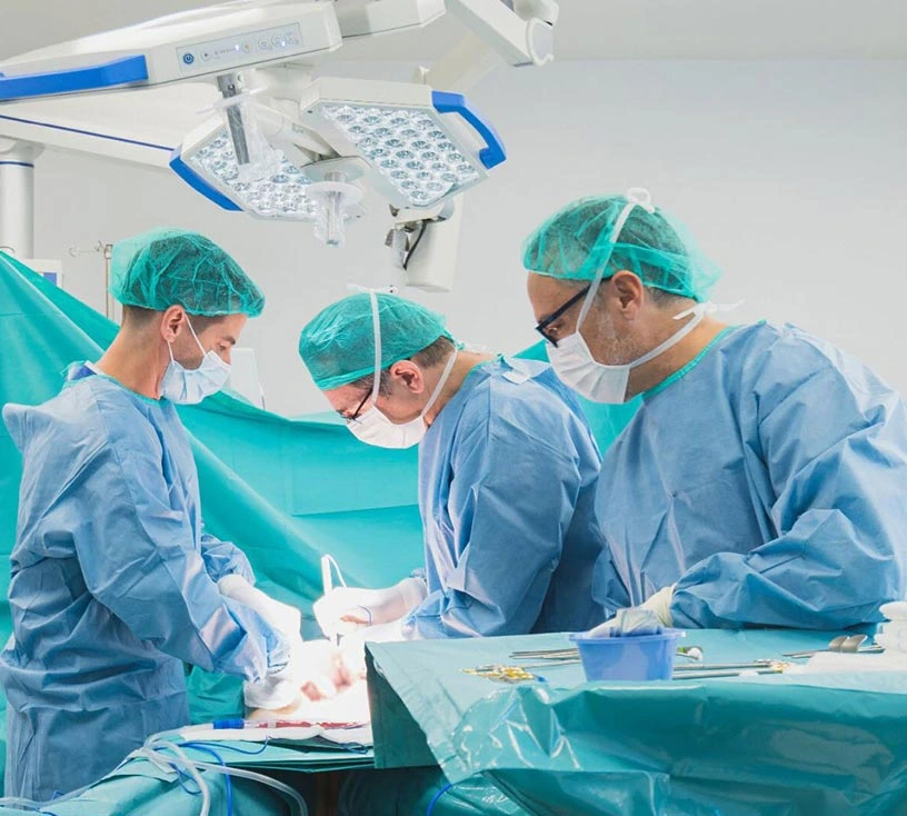
Liver metastases are considered operable when all visible disease can be removed while leaving a liver that works. Today the decision does not depend on the number or size of the lesions, as was believed decades ago, but on three conditions: that a complete resection with a clear margin can be achieved, or a partial resection combined with ablation; that the liver left behind is sufficient and has good blood supply and drainage; and that the biology of the tumor makes surgery reasonable. Many patients who were told they could not be operated on turn out to be operable after an assessment in a specialized hepatobiliary unit.
What it means today for liver metastases to be operable
For years the surgical indication relied on numerical criteria: no more than three lesions, none larger than five centimeters, a margin of at least one centimeter. Those limits excluded many patients who today are operated on with good results and for whom surgery means living longer.
The conceptual change is that resectability is no longer defined by what is removed, but by what remains. The question stopped being how many tumors there are and became whether the remaining liver will be enough to support the patient.
This shift explains why two teams can reach different conclusions from the same CT scan. It is not a disagreement on medical judgment: the assessment depends on the technical resources available and on the team's experience in complex liver surgery.
Technical resectability and oncological resectability
They are two different questions, and the answer to both has to be yes.
Technical resectability asks whether the operation can be done safely. It relies on anatomical and functional criteria:
- Complete resection. It must be possible to remove all visible disease. A one-centimeter margin is no longer required: today a smaller margin is accepted as long as it is negative. In specific cases even a margin in contact with a blood vessel of the liver is accepted, and this has greatly increased the number of patients who can be operated on.
- Sufficient remaining liver volume. This is the criterion that has gained the most weight. It is estimated with CT volumetry and expressed as a percentage of the total liver. The required threshold varies with the condition of the liver: it is higher when the liver has received prolonged chemotherapy or shows steatosis or fibrosis.
- Vascular and biliary preservation. The remaining liver must keep its arterial and portal inflow, outflow through at least one hepatic vein, and biliary drainage. A small lesion next to a hepatic vein can affect operability more than several large peripheral lesions.
Oncological resectability asks whether operating makes sense for that particular patient. What matters here is how the tumor behaves, the interval between the primary tumor and the appearance of the metastases, the response to previous systemic treatment, tumor markers and the molecular profile. A tumor that progresses during chemotherapy behaves differently from one that responds, and that information is as relevant to the decision as the imaging.
Criteria that no longer rule out surgery
Three situations that twenty years ago closed the door to surgery and today do not, on their own:
The number of lesions. No figure automatically excludes surgery. What matters is distribution: five lesions grouped in one lobe may be easier to treat than two located where removing them would sacrifice too much liver.
Involvement of both lobes. Lesions in both lobes are not a contraindication. Two-stage strategies and combinations of resection and ablation were designed precisely for these cases.
Disease outside the liver. It is no longer an absolute contraindication. Limited, resectable lung involvement, or peritoneal disease suitable for specific treatment, can be addressed within an overall strategy.
When the remaining liver is not enough: how it is increased
A patient may not be operable today and become operable in a few weeks. These are the strategies that turn an initially unresectable situation into an operable one.
Conversion chemotherapy. It is not the same as palliative chemotherapy: the aim is to shrink the lesions until they can be removed. It requires repeat imaging at short intervals and close coordination between oncology and surgery, because the window in which the tumor is resectable can be short. Continuing it for too long carries two risks: liver toxicity from the treatment itself, and lesions that stop showing on imaging without having really disappeared, which makes them hard to find in the operating room. Today there are new systemic therapies and strategies that reduce the size and number of metastases and make surgery possible afterwards.
Portal vein embolization. The portal branch on the side to be removed is blocked so that the side that will remain grows. After a few weeks the volume is measured again; if it has grown enough, surgery goes ahead.
Two-stage hepatectomy. The future remaining liver is cleared first, the liver is allowed to regenerate, and the resection is completed in a second operation.
Ablation combined with resection. For small, deep lesions whose removal would cost too much liver, resection of some lesions can be combined with ablation of others.
Hepatic arterial infusion chemotherapy. The treatment is given directly into the hepatic artery, which allows higher concentrations in the liver with less effect on the rest of the body. It is an option in selected cases of disease limited to the liver and increases the effectiveness of treatment on the metastases. We explain it in the article on hepatic arterial infusion chemotherapy.
Liver transplantation. In recent years, liver transplantation has been validated for patients with liver metastases of colorectal origin who meet a set of strict criteria, with very good results. This means that patients whose metastases cannot be removed may benefit from a treatment that extends their survival.
Not all liver metastases follow the same rules
Much of the above comes from experience with metastases of colorectal origin, which are the most common and the most studied. The criteria change with the primary tumor: in neuroendocrine tumors the surgical indication follows a different logic, and in metastases of other origins surgery is considered in more specific scenarios.
That is why an operability assessment requires knowing the primary tumor, its histology and its molecular profile. An opinion based only on liver imaging is incomplete.
Who decides, and why it matters where the decision is made
The surgical indication for liver metastases is not made by a single professional. It is decided by a multidisciplinary board, with hepatobiliary surgery, medical oncology, radiology and pathology assessing the case together.
This has a practical consequence worth understanding: "not operable" sometimes means "not operable with the resources of this center". Complex liver surgery (extended resections, vascular reconstruction, two-stage strategies) is concentrated in units that treat many of these cases. A center without that capacity may reasonably rule out an operation that a specialized unit would consider.
If you have been told your case is not operable, asking for a second assessment in a hepatobiliary unit does not question anyone: it is the logical next step. For that assessment you need the imaging studies in their original format (the images, not just the report), the pathology report of the primary tumor, details of the treatments received with their dates, and the molecular profile if it has been done.
The surgical techniques are described in liver resections, and prognosis figures in life expectancy with liver metastases.
Frequently asked questions
How many liver metastases can be operated on?
There is no limit. What determines operability is the distribution of the lesions and the volume of healthy liver that will remain afterwards, not how many there are.
Can surgery be done if there are lesions in both lobes?
Yes. Involvement of both lobes does not rule out surgery on its own: two-stage strategies and combinations of resection and ablation were designed for these cases.
I have been told I am inoperable. Can that change?
It can. Some patients who are initially unresectable become resectable after conversion chemotherapy or after techniques that increase the volume of the remaining liver. The assessment also depends on the team's experience, so a second opinion in a hepatobiliary unit is justified.
How long does it take to know if I am a candidate?
With the imaging studies and reports available, the initial assessment takes days. If further tests are needed or the case has to be presented to the board, it takes a little longer.
This information is for general guidance and does not replace an assessment by your medical team.