
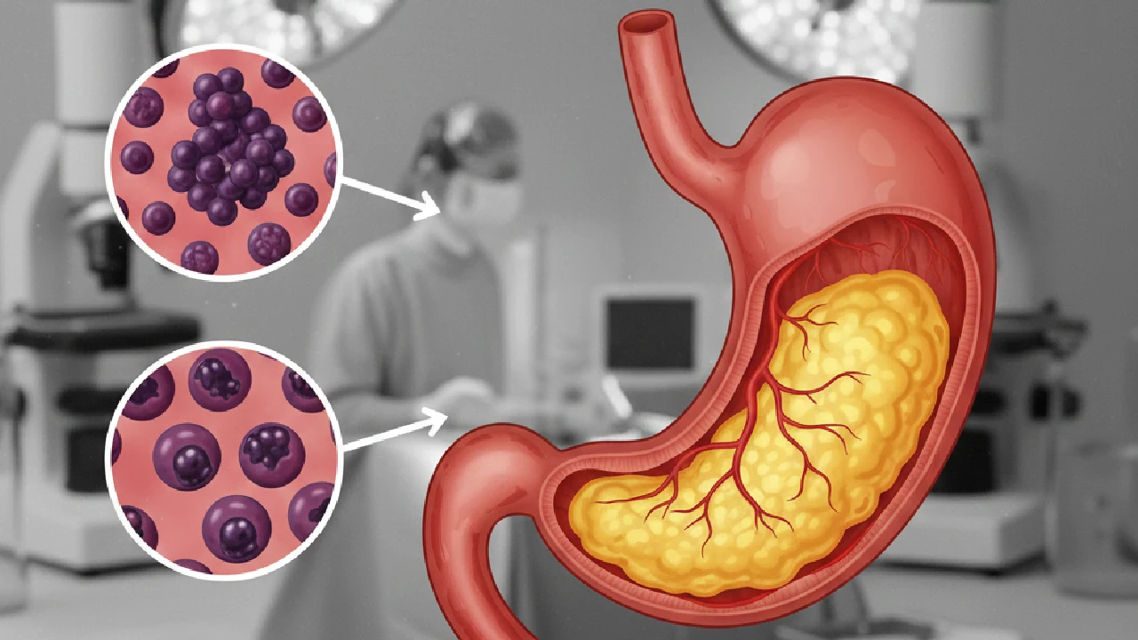
What is stomach cancer?
Stomach cancer (also called gastric cancer) is a malignant tumour that starts in the cells lining the inside of the stomach. It is the fifth most common cancer worldwide, with the highest incidence in Asian countries.
The most common histological type is adenocarcinoma (90-95% of cases), which can be intestinal type (associated with chronic gastritis) or diffuse type (more aggressive, associated with CDH1 mutations).
Causes and risk factors
Helicobacter pylori infection: The main risk factor. It causes chronic gastritis that can progress to intestinal metaplasia, dysplasia and cancer. It is responsible for 60-80% of cases.
Dietary factors: High intake of salt, smoked foods, salt-preserved foods, nitrates and nitrites. Low intake of fresh fruit and vegetables.
Smoking: Doubles the risk of gastric cancer.
Family history: Hereditary syndromes such as hereditary diffuse gastric cancer (CDH1 mutation), Lynch syndrome, familial adenomatous polyposis.
Chronic atrophic gastritis: Especially when associated with pernicious anaemia.
Previous gastric surgery: Partial gastrectomy increases the long-term risk.
Older age: Risk increases after the age of 60.
Symptoms of stomach cancer
In the early stages it usually causes no symptoms, which is why screening matters in at-risk populations. When symptoms appear, they can include:
• Persistent dyspepsia: Digestive discomfort that does not improve with the usual treatment
• Early satiety: Feeling full quickly when eating
• Unintentional weight loss and loss of appetite (anorexia)
• Epigastric pain, continuous or intermittent
• Nausea and vomiting, which may contain blood in advanced stages
• Iron-deficiency anaemia from chronic hidden bleeding
• Dysphagia: Difficulty swallowing, especially in tumours of the cardia or the gastro-oesophageal junction
• Melaena: Black stools from gastrointestinal bleeding
Diagnosis
Gastroscopy (upper gastrointestinal endoscopy): The gold-standard test. It shows the tumour directly, allows multiple biopsies to be taken for histological study and assesses local extent.
Contrast-enhanced chest and abdominal CT: For staging and to assess infiltration of neighbouring organs, lymph nodes and distant metastases.
Endoscopic ultrasound: Assesses the depth of invasion in the stomach wall (T stage) and regional lymph node involvement more accurately than CT.
PET-CT: In selected cases, to detect hidden metastatic disease before major surgery.
Diagnostic laparoscopy: In locally advanced tumours, to rule out peritoneal carcinomatosis not visible on CT (present in 20-30% of advanced cases).
Treatment: a multidisciplinary approach
Surgery: The only potentially curative treatment. The type of surgery depends on the location and extent of the tumour:
• Subtotal gastrectomy: For tumours of the distal gastric antrum
• Total gastrectomy: For tumours of the gastric body, fundus or cardia, or multicentric tumours
• D2 lymphadenectomy: Extensive lymph node resection (at least 15 nodes), needed for correct staging and for oncological control
At Quenet Torrent Institute we perform gastrectomies using robotic surgery and laparoscopy, with better postoperative recovery, less pain and shorter hospital stays, while keeping the same oncological results as open surgery.
Perioperative chemotherapy: In locally advanced tumours (stage II-III), chemotherapy before (neoadjuvant) and after (adjuvant) surgery significantly improves survival. Regimens: FLOT (5-fluorouracil, leucovorin, oxaliplatin, docetaxel) or ECF/EOX.
Radiotherapy: Mainly in tumours of the gastro-oesophageal junction, combined with chemotherapy.
Targeted therapies and immunotherapy: In advanced or metastatic disease:
• Trastuzumab: For HER2-positive tumours (15-20% of cases)
• Pembrolizumab: Immunotherapy in tumours with high microsatellite instability or high PD-L1 expression
• Ramucirumab: Anti-VEGFR2 antibody in second line
Prognosis
The prognosis depends mainly on the stage at diagnosis:
• Stage I (tumour limited to the mucosa/submucosa): 5-year survival >80%
• Stage II (muscle invasion, few lymph nodes): Survival 50-70%
• Stage III (serosal invasion, multiple lymph nodes): Survival 20-40%
• Stage IV (metastatic): Survival <10%
That is why early detection matters so much. In Japan and Korea, where there is population screening, 50% of cases are diagnosed at stage I, with high cure rates. In Spain, only 15-20% are diagnosed at early stages.
Prevention
H. pylori eradication: Antibiotic treatment of Helicobacter pylori infection significantly reduces the risk of gastric cancer. A "test and treat" approach is recommended in people with a family history.
Healthy diet: High in fresh fruit and vegetables, less salt, and avoiding smoked and salt-preserved foods.
Stopping smoking: Quitting smoking reduces the risk of gastric cancer and many other cancers.
Endoscopic surveillance: In patients with atrophic gastritis, extensive intestinal metaplasia, dysplasia or a first-degree family history of gastric cancer.
Prophylactic gastrectomy: In carriers of a CDH1 mutation with hereditary diffuse gastric cancer (after genetic counselling).
When gastric cancer has spread to the peritoneum
This is the scenario in which the door to surgery is most often closed, and it deserves some nuance. Stomach cancer often spreads to the peritoneum, and that diagnosis is frequently communicated simply as terminal disease.
The reality is more nuanced. In selected patients, with limited peritoneal disease and good general health, there are options that are not only palliative:
• Cytoreduction with HIPEC in peritoneal carcinomatosis of gastric origin: selection here is stricter than for other origins, with low PCI thresholds, but in the right group it changes how the disease progresses.
• PIPAC: pressurised aerosol chemotherapy when complete cytoreduction cannot be achieved. It is a repeatable procedure with quick recovery, aimed at controlling the disease and the ascites.
• Staging laparoscopy with peritoneal cytology: before ruling anyone out, it is worth knowing exactly how much peritoneal disease there is. An isolated positive cytology is not the same as established carcinomatosis.
The difference between one case and another cannot be seen in a CT report. It is seen by looking inside.
Have you been told that your gastric cancer can no longer be operated on because of peritoneal involvement? This is one of the situations we assess most often, and the answer depends on accurate staging. We review the case and tell you frankly whether there is a surgical option. Request an assessment.